
Switch to Gallery View
Image and Video Gallery
This is a searchable collection of scientific photos, illustrations, and videos. The images and videos in this gallery are licensed under Creative Commons Attribution Non-Commercial ShareAlike 3.0. This license lets you remix, tweak, and build upon this work non-commercially, as long as you credit and license your new creations under identical terms.

Plasma-Derived Membrane Vesicles
5887
This fiery image doesn’t come from inside a bubbling volcano. Instead, it shows animal cells caught in the act of making bubbles, or blebbing. Jeanne Stachowiak, University of Texas at Austin View Media
Optic nerve astrocytes
5852
Astrocytes in the cross section of a human optic nerve head Tom Deerinck and Keunyoung (“Christine”) Kim, NCMIR View Media
Cryo-ET cell cross-section visualizing insulin vesicles
6607
On the left, a cross-section slice of a rat pancreas cell captured using cryo-electron tomography (cryo-ET). On the right, a color-coded, 3D version of the image highlighting cell structures. Xianjun Zhang, University of Southern California. View Media
Bovine milk alpha-lactalbumin (2)
2404
Crystals of bovine milk alpha-lactalbumin protein created for X-ray crystallography, which can reveal detailed, three-dimensional protein structures. Alex McPherson, University of California, Irvine View Media
Protein purification robot
2375
Irina Dementieva, a biochemist, and Youngchang Kim, a biophysicist and crystallographer, work with the first robot of its type in the U.S. to automate protein purification. Midwest Center for Structural Genomics View Media
Fungal lipase (2)
2411
Crystals of fungal lipase protein created for X-ray crystallography, which can reveal detailed, three-dimensional protein structures. Alex McPherson, University of California, Irvine View Media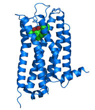
Dopamine D3 receptor
3363
The receptor is shown bound to an antagonist, eticlopride Raymond Stevens, The Scripps Research Institute View Media
Automated crystal screening system
2362
Automated crystal screening systems such as the one shown here are becoming a common feature at synchrotron and other facilities where high-throughput crystal structure determination is being carried Southeast Collaboratory for Structural Genomics View Media
Hsp33 Heat Shock Protein Inactive to Active
3402
When the heat shock protein hsp33 is folded, it is inactive and contains a zinc ion, stabilizing the redox sensitive domain (orange). Dana Reichmann, University of Michigan View Media
Arachnoidiscus diatom
6902
An Arachnoidiscus diatom with a diameter of 190µm. Michael Shribak, Marine Biological Laboratory/University of Chicago. View Media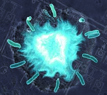
Supernova bacteria
2725
Bacteria engineered to act as genetic clocks flash in synchrony. Here, a "supernova" burst in a colony of coupled genetic clocks just after reaching critical cell density. Jeff Hasty, UCSD View Media
Body toxins (with labels)
2497
Body organs such as the liver and kidneys process chemicals and toxins. These "target" organs are susceptible to damage caused by these substances. Crabtree + Company View Media
Anchor cell in basement membrane
2707
An anchor cell (red) pushes through the basement membrane (green) that surrounds it. Elliott Hagedorn, Duke University. View Media
Breast cancer cells change migration phenotypes
6986
Cancer cells can change their migration phenotype, which includes their shape and the way that they move to invade different tissues. Bo Sun, Oregon State University. View Media
Lily mitosis 11
1011
A light microscope image of cells from the endosperm of an African globe lily (Scadoxus katherinae). This is one frame of a time-lapse sequence that shows cell division in action. Andrew S. Bajer, University of Oregon, Eugene View Media
tRNA splicing enzyme endonuclease in humans
2351
An NMR solution structure model of the transfer RNA splicing enzyme endonuclease in humans (subunit Sen15). This represents the first structure of a eukaryotic tRNA splicing endonuclease subunit. Center for Eukaryotic Structural Genomics, PSI View Media
HeLa cell undergoing division into two daughter cells
6520
Here, a human HeLa cell (a type of immortal cell line used in laboratory experiments) is undergoing cell division. Dylan T. Burnette, Ph.D., Vanderbilt University School of Medicine. View Media
Magnetic Janus particle activating a T cell
6800
A Janus particle being used to activate a T cell, a type of immune cell. Yan Yu, Indiana University, Bloomington. View Media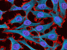
HeLa cells
3521
Multiphoton fluorescence image of HeLa cells stained with the actin binding toxin phalloidin (red), microtubules (cyan) and cell nuclei (blue). Nikon RTS2000MP custom laser scanning microscope. National Center for Microscopy and Imaging Research (NCMIR) View Media
Fruitful dyes
2317
These colorful, computer-generated ribbons show the backbone of a molecule that glows a fluorescent red. Roger Y. Tsien, University of California, San Diego View Media
Yeast cells responding to a glucose shortage
6772
These yeast cells were exposed to a glucose (sugar) shortage. Mike Henne, University of Texas Southwestern Medical Center. View Media
Mouse colon with gut bacteria
3566
A section of mouse colon with gut bacteria (center, in green) residing within a protective pocket. Sarkis K. Mazmanian, California Institute of Technology View Media
Induced stem cells from adult skin 01
2603
These cells are induced stem cells made from human adult skin cells that were genetically reprogrammed to mimic embryonic stem cells. James Thomson, University of Wisconsin-Madison View Media
Floral pattern in a mixture of two bacterial species, Acinetobacter baylyi and Escherichia coli, grown on a semi-solid agar for 48 hours (photo 1)
6553
Floral pattern emerging as two bacterial species, motile Acinetobacter baylyi (red) and non-motile Escherichia coli (green), are grown together for 48 hours on 1% agar surface from a sma L. Xiong et al, eLife 2020;9: e48885 View Media
Antitoxin GhoS (Illustration 1)
3427
Structure of the bacterial antitoxin protein GhoS. GhoS inhibits the production of a bacterial toxin, GhoT, which can contribute to antibiotic resistance. Rebecca Page and Wolfgang Peti, Brown University and Thomas K. Wood, Pennsylvania State University View Media
Pathways – Bacteria vs. Viruses: What's the Difference?
6597
Learn about how bacteria and viruses differ, how they each can make you sick, and how they can or cannot be treated. National Institute of General Medical Sciences View Media
Protein clumping in zinc-deficient yeast cells
3550
The green spots in this image are clumps of protein inside yeast cells that are deficient in both zinc and a protein called Tsa1 that prevents clumping. Colin MacDiarmid and David Eide, University of Wisconsin--Madison View Media
Caulobacter
3262
A study using Caulobacter crescentus showed that some bacteria use just-in-time processing, much like that used in industrial delivery, to make the glue that allows them to attach to surfaces, Yves Brun, Indiana University View Media
Bacterial ribosome assembly
6578
3D reconstructions of two stages in the assembly of the bacterial ribosome created from time-resolved cryo-electron microscopy images. Ribosomes translate genetic instructions into proteins. Joachim Frank, Columbia University. View Media
Staphylococcus aureus in the porous coating of a femoral hip stem
6804
Staphylococcus aureus bacteria (blue) on the porous coating of a femoral hip stem used in hip replacement surgery. Paul Stoodley, The Ohio State University. View Media
Cone snail shell
2576
A shell from the venomous cone snail Conus omaria, which lives in the Pacific and Indian oceans and eats other snails. Kerry Matz, University of Utah View Media
Highlighted cells
2429
The cytoskeleton (green) and DNA (purple) are highlighed in these cells by immunofluorescence. Torsten Wittmann, Scripps Research Institute View Media
Wound healing in process
3498
Wound healing requires the action of stem cells. Hermann Steller, Rockefeller University View Media
Regenerating lizard tail
6968
The interior of a regenerating lizard tail 14 days after the original tail was amputated. Thomas Lozito, University of Southern California. View Media
Floral pattern in a mixture of two bacterial species, Acinetobacter baylyi and Escherichia coli, grown on a semi-solid agar for 72 hour
6556
Floral pattern emerging as two bacterial species, motile Acinetobacter baylyi and non-motile Escherichia coli (green), are grown together for 72 hours on 0.5% agar surface from a small i L. Xiong et al, eLife 2020;9: e48885 View Media
2-D NMR
2299
A two-dimensional NMR spectrum of a protein, in this case a 2D 1H-15N HSQC NMR spectrum of a 228 amino acid DNA/RNA-binding protein. Dr. Xiaolian Gao's laboratory at the University of Houston View Media
Group of fluorescent C. elegans showing muscle and ribosomal protein
6582
Three C. elegans, tiny roundworms, with a ribosomal protein glowing red and muscle fibers glowing green. Researchers used these worms to study a molecular pathway that affects aging. Jarod Rollins, Mount Desert Island Biological Laboratory. View Media
Pig alpha amylase
2412
Crystals of porcine alpha amylase protein created for X-ray crystallography, which can reveal detailed, three-dimensional protein structures. Alex McPherson, University of California, Irvine View Media
Telomeres on outer edge of nucleus during cell division
3484
New research shows telomeres moving to the outer edge of the nucleus after cell division, suggesting these caps that protect chromosomes also may play a role in organizing DNA. Laure Crabbe, Jamie Kasuboski and James Fitzpatrick, Salk Institute for Biological Studies View Media
Mouse cerebellum in pink and blue
5800
The cerebellum is the brain's locomotion control center. Found at the base of your brain, the cerebellum is a single layer of tissue with deep folds like an accordion. National Center for Microscopy and Imaging Research (NCMIR) View Media
Anti-tumor drug ecteinascidin 743 (ET-743), structure without hydrogens 01
2794
Ecteinascidin 743 (ET-743, brand name Yondelis), was discovered and isolated from a sea squirt, Ecteinascidia turbinata, by NIGMS grantee Kenneth Rinehart at the University of Illinois. Timothy Jamison, Massachusetts Institute of Technology View Media
Computer model of cell membrane
2636
A computer model of the cell membrane, where the plasma membrane is red, endoplasmic reticulum is yellow, and mitochondria are blue. Bridget Wilson, University of New Mexico View Media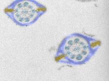
Mouse sperm sections
1191
This transmission electron micrograph shows sections of mouse sperm tails, or flagella. Tina Weatherby Carvalho, University of Hawaii at Manoa View Media
Actin filaments bundled around the dynamin helical polymer
6571
Multiple actin filaments (magenta) are organized around a dynamin helical polymer (rainbow colored) in this model derived from cryo-electron tomography. Elizabeth Chen, University of Texas Southwestern Medical Center. View Media
Glucose and sucrose
2500
Glucose (top) and sucrose (bottom) are sugars made of carbon, hydrogen, and oxygen atoms. Carbohydrates include simple sugars like these and are the main source of energy for the human body. Crabtree + Company View Media
Bacterial spore
2752
A spore from the bacterium Bacillus subtilis shows four outer layers that protect the cell from harsh environmental conditions. Patrick Eichenberger, New York University View Media
Protein rv2844 from M. tuberculosis
2343
This crystal structure shows a conserved hypothetical protein from Mycobacterium tuberculosis. Only 12 other proteins share its sequence homology, and none has a known function. Integrated Center for Structure and Function Innovation View Media
Anti-tumor drug ecteinascidin 743 (ET-743) with hydrogens 02
2791
Ecteinascidin 743 (ET-743, brand name Yondelis), was discovered and isolated from a sea squirt, Ecteinascidia turbinata, by NIGMS grantee Kenneth Rinehart at the University of Illinois. Timothy Jamison, Massachusetts Institute of Technology View Media
CRISPR Illustration
7036
This illustration shows, in simplified terms, how the CRISPR-Cas9 system can be used as a gene-editing tool. National Institute of General Medical Sciences. View Media
Bacterial cells aggregating above the light organ of the Hawaiian bobtail squid
7018
A light organ (~0.5 mm across) of a juvenile Hawaiian bobtail squid, Euprymna scolopes. Margaret J. McFall-Ngai, Carnegie Institution for Science/California Institute of Technology, and Edward G. Ruby, California Institute of Technology. View Media


